Research Achievement
2019
본 연구단의 중점 연구분야 중 하나는 상호작용이 강한 이차원 물질(TaS2, IrTe2, Ta2NiSe5 등)에서의 이종계면 또는 도메인월(위상여기)이다.
올해는 본 연구단이 2016년 이후 높은 수준의 선도적인 성과를 내고 있는 1T-TaS2 계의 도메인월에 대한 연구를 확장 심화시켰다. 본 물질이 가지는 독특한 Mott-전하밀도파 상태에서의 도메인월은 본 연구단이 2016년 및 2017년의 Nature Communications 논문에서 저온에서 발생하는 도메인월의 구조 및 그 파급효과 등을 세계최초로 보고하였으나, 해당 도메인월 구조들은 절연체적인 성질을 가지고 있어, 도메인월이 중요할 것으로 생각되는 1T-TaS2에서의 초전도 현상과의 연결고리가 무엇인지 중대한 의문으로 남았다.
본 연구에서는 초전도현상과 직접 관련이 있는 도메인월 네트워크가 형성되어 있는 nearly commensurate CDW 상의 도메인월 구조를 세계 최초로 원자분해능으로 직접 관찰하는데 성공하였으며, 아울러 이러한 원자구조를 전자구조계산을 통하여 규명할 수 있었다.
그 결과, 자연적으로 여기되는 고온상의 도메인월 구조는 저온의 구조와는 다른 원자구조를 가지며, 이 구조가 금속적인 전자상태의 네트워크를 가진다는 사실을 밝혀낼 수 있었다. 이 결과로부터 1T-TaS2 계에서 나타나는 초전도 현상을 자연스럽게 설명할 수 있는 길이 열리게 되었다.
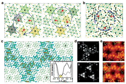
Figure 2. Structure models (a, b), local density of states (c) and STM images (d, e) of domain walls in the chare density wave state of 1T-TaS2.
When two periodic potentials compete in materials, one may adopt the other, which straightforwardly generates topological defects.
Of particular interest are domain walls in charge-, dipole-, and spin- ordered systems, which govern macroscopic properties and important functionality. However, detailed atomic and electronic structures of domain walls have often been uncertain and the microscopic mechanism of their functionality has been elusive.
Here, we clarify the complete atomic and electronic structures of the domain wall network, a honeycomb network connected by Z3 vortices, in the nearly commensurate Mott charge-density wave (CDW) phase of 1T-TaS2. Scanning tunneling microscopy resolves characteristic charge orders within domain walls and their vortices.
Density functional theory calculations disclose their unique atomic relaxations and the metallic in-gap states confined tightly therein. A generic theory is constructed, which connects this emergent honeycomb network of conducting electrons to the enhanced superconductivity.
2H-NbSe2는 전자-포논 상호작용 강한 대표적인 이차원물질로서 상호작용을 통한 전하밀도파와 초전도의 발생 및 공존이 오랫동안 알려져왔다. 특히 전하밀도파의 경우 강한 전자-포논 상호작용이 중요하다고 잘 알려졌으나, 저온의 기저상태가 incommensurate 상태를 보이는 이유가 그 동안 베일에 쌓여있었다.
최근 전하밀도파와 초전도의 공존이 전하밀도파의 incommensuration과 관련이 깊다는 것이 점차 알려지고 있어, 2H-NbSe2과 같은 대표적인 물질계에서의 incommensuration의 원인을 밝히는 것은 중요한 과제로 대두되었다.
본 연구에서는 주사터널현미경을 활용하여 해당 이차원 물질의 전하밀도파 상을 이전의 연구들과는 다른 정밀도로 측정한 결과, 이전의 연구들이 발견하지 못했던, 두가지 서로 다른 전하밀도파 상이 공존하는 양상을 세계 최초로 발견하였다. 더 나아가 이러한 구조 각각에 대한 전자구조 계산, 그리고 그들이 공존하여 만드는 계면구조에 대한 자세한 실험과 계산을 통하여, 이러한 두 가지 다른 구조의 공존이 incommensuration의 기원임을 밝힐 수 있었다. 이는 지난 50여 년간 미스테리였던 2H-NbSe2에서의 전하밀도파 incommensuration의 원인을 밝히는데 그치지 않고, incommensuration이 일반적인 수평이동형 도메인월이 아닌 완전히 다른 메카니즘을 통해서도 만들어 질 수 있음을 보여, 저차원계 물리연구의 지평을 넓혔다 할 수 있다.
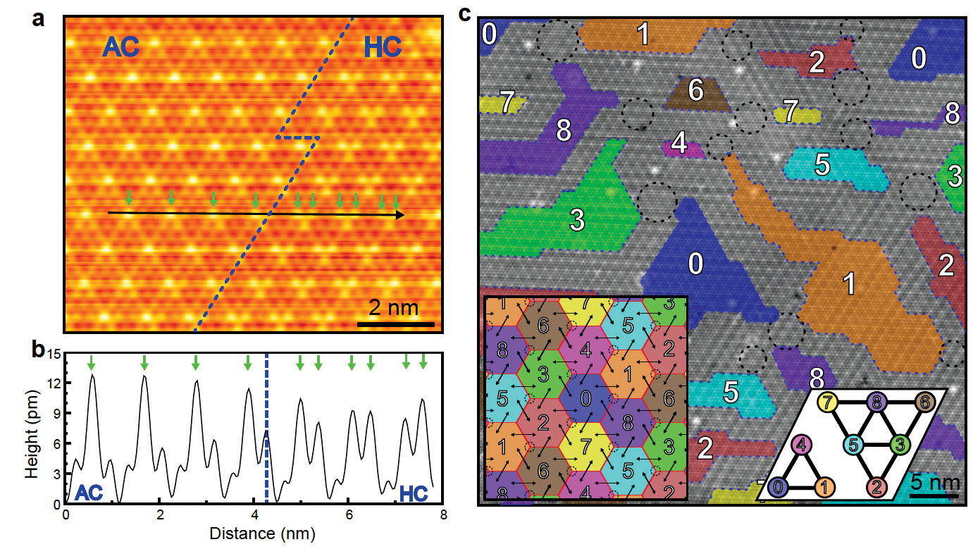
Figure 1. Scanning tunneling microscopy images of coexisting charge density wave structures in 2H-NbSe2.
Despite decades of studies of the charge density wave (CDW) of 2H-NbSe2, the origin of its incommensurate CDW ground state has not been understood. We discover that the CDW of 2H-NbSe2 is composed of two different, energetically competing, structures.
The lateral heterostructures of two CDWs are entangled as topological excitations, which give rise to a CDW phase shift and the incommensuration without a conventional domain wall.
A partially melted network of topological excitations and their vertices explain an unusual landscape of domains. The unconventional topological role of competing phases disclosed here can be widely applied to various incommensuration or phase coexistence phenomena in materials.
Ta2NiSe5 물질은 본 연구단에서 합성된 시료로서, 강한 Coulomb 상호작용에 의하여 높은 온도에서 엑시톤 절연체가 되는 매우 특이한 물성을 지니는 물질로 최근 주목을 받고 있다.
본 연구단은 이전 연구에서 이 물질의 엑시톤 갭을 분명하게 측정하였으며, 이러한 에너지 갭이 전자와 홀 밴드 사이의 강한 hybridization에 기인한다는 것을 실험과 이론적으로 밝혔다.
본 연구에서는 해당 시료에 강한 전기장을 인가하여 엑시톤 상호작용 즉, 전자와 홀밴드 hybridization의 조절 가능성을 검토하였다.
매우 강한 전기장을 시료 표면에 인가하기 위하여 표면에 알칼리금속 이온을 흡착시켰고, 흡착량을 조절하여 인가된 평균적 전기장의 세기를 조절하는 잘 알려진 표면 도핑 방법을 활용하였다. 그 결과 아래의 전자밴드구조 측정에서 보여주듯, 전자밴드와 홀밴드 사이의 hybridization의 정도에 해당되는 엑시톤 절연체상의 밴드의 곡률이 인가된 전기장에 비례하여 증가함을 알 수 있었다. 이는 이론적으로 예측되는 엑시톤 절연체에 대한 전기장 인가 효과와 잘 일치하였다.
이 실험을 통하여 이차원 물질계에 존재하는 강한 상호작용을 제어가능한 외부적인 요소 (이 경우 전기장)를 통하여 조절할 수 있음이 밝혀졌으며, 이는 이러한 강한 상호작용에 기반한 다양한 기능성 물질과 소자의 응용에 활용될 수 있는 방법이라 할 수 있다. 특히 최근 주목을 받고 있는 이차원계의 엑시톤 소자와 관련하여 시사점이 큰 연구라고 할 수 있다.
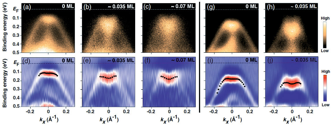
Figure 3. Experimentally measured (by angle resolved photoelectron spectroscopy) band structures of Ta2NiSe5 at different coverages of K adsorbates on the surface, showing the change of excitonic coupling in the valence band top.
We demonstrate that the excitonic insulator ground state of Ta2NiSe5 can be electrically controlled by electropositive surface adsorbates. Our studies utilizing angle-resolved photoemission spectroscopy reveal intriguing wave-vector-dependent deformations of the characteristic flattop valence band of this material upon potassium adsorption. The observed band deformation indicates a reduction of the single-particle band gap due to the Stark effect near the surface. The present study provides the foundation for the electrical tuning of the many-body quantum states in excitonic insulators.
새로운 반도체 물질로 주목받는 원자층 이차원 반도체 전이금속 칼코겐 화합물은 높은 접촉 저항을 갖는 것이 심각한 기술적 난제로 알려져 있어 이를 해결하는 것이 매우 중요하다. 본 연구에서는 이차원 전이금속 칼코겐 화합물이 오비탈 중첩의 차이에 따라 다양한 구조와 전기적 물성을 갖는 다형체임을 이용하여, 금속성을 갖는 WTe2와 반도체성을 갖는 WSe2의 단일층 이종 수직 접합 합성을 통해 “반데르 발스 에피 접합”이라는 새로운 원자층 반도체-금속 접합을 최초로 구현하였다. 즉 연속적인 화학 기상 증착 공정을 통하여 합성된 이종 접합은 원자수준에서 깨끗한 계면을 가지고, 또한 격자 결맞음이 유지되는 고품위의 금속-반도체 접합 구조체가 된다. 이러한 반데르 발스 에피 접합은 일반 금속 접합보다 2배 이상 낮은 저항 접합을 가지며 이를 통해 100배 이상의 높은 전하 이동도를 확보할 수 있음을 보였다.
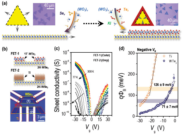
Figure 4. (a) Sequential growth scheme for heteroepitaxial stacking of 2H-WSe2 and 1T’-WTe2 monolayers (MLs). Inset: optical microscope images of ML WSe2 (left) and bilayer WTe2/WSe2 (right). (b) Illustration of our device scheme for comparison of vdW epitaxial metal (1T’-WTe2) contact (FET-1) with planar metal (Ti) contact (FET-2) and optical microscope image of vdW epitaxial contact devices. (c) Temperature-dependent Vg modulation of sheet conductivity (σ) at Vb = 50 mV for FET-1 (color scale) and FET-2 (gray scale). (d) Built-in potential energy (qΦbi) as a function of Vg for FET-1 (1T’-WTe2 contact, navy), FET-2 (Ti contact, orange).
In the atomic layer two-dimensional semiconductors, transition-metal chalcogenides, the very high contact resistance is important technical challenges to be solved. Some of transition-metal chalcogenides are known to exist in various polymorphs with different electronic phases as metals or semiconductors.
In this study, we for the first time demonstrated a new atomic layer semiconductor-metal junction called as “epitaxial van der Waals contact” via vertical integration of metallic WTe2 and semiconducting WSe2 by sequential MOCVD processes. These synthetic heterostructures form high-quality metal-semiconductor junctions with atomically cleaninterface and lattice coherency.
Our epitaxial van der Waals contact realized much lower contact barrier potentials than conventional metal contacts, resulting in the 100 times larger charge carrier mobility in field-effect transistors.
원자층 두께의 반도체를 기판에서 분리한다면 유연한 원자막으로 존재할 수 있는데 이를 “멤브레인 반도체”라고 부를 수 있다. 지금까지 2차원 반도체는 통상적으로 평면 형태로만 합성되었는데, 이는 극도로 얇은 원자층 두께 특성으로 인하여 굴곡 모양의 격자 형성이 불완전했기 때문이다.
본 연구에서는 결정 성장 속도를 원자수준에서 제어 가능한 MOCVD 기법을 개발하여 나노 크기의 굴곡 (10 nm 크기의 바늘 모양 돌기 어레이) 위에 2차원 반도체인 MoS2를 원자층 3차원 구조로 성장하는데 성공하였다. 또한 성장된 3차원 구조체는 간단히 물에 담가 기판으로부터 떼어낼 수 있어, 멤브레인 반도체 형태로 구현할 수 있다.
이러한 특성은 원자층 반도체를 원하는 제3의 기판위로 전사 가능함을 예시하며, 따라서 이러한 멤브레인 반도체는 플라스틱, 인체 피부 등 다양한 patchable electronics 형태로 개발이 가능한 물질 플랫폼으로 활용될 수 있다.
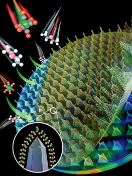
Figure 5. A schematic illustration depicting the conformal growth and seperation of atomically thin semiconductor films in a three-dimensional membrane form with monolayer precision.
When an atomically thin two-dimensional (2D) semiconductor film is delaminated from silicon wafers, it becomes a flexible membrane at the atomic scales, called “semiconductor membranes”.
Until now, 2D semiconductors have been deposited on planar forms, because the lattice formation on the curved surface is incomplete due to the extremely thin atomic layer thickness characteristics. In this study, we have succeeded in uniformly growing three-dimensional atomic-layer MoS2 on array of sharp needles (tip radius = 10 nm) via MOCVD methods, which enables the crystal growth rate at the atomic level. Our 3D atomic-layer semiconductors can be easily detachable from the wafer by soaking them in water, realizing the form of semiconductor membrane. These exemplify that an atomic-layer semiconductors can be transferred onto any desired substrates, thus semiconductor membranes can be utilized as new material platforms for patchable electronics on various surfaces (e.g., plastic and human skin) just like a Post-it.
물질의 전자구조를 직접적으로 측정할 수 있는 각도분해 광전자 분광법은 그 물성이 전통적 밴드 계산만으로는 설명되지 않는 강상관계 물질 및 차원과 위상에 의해 물성이 결정되는 위상부도체, Weyl 반금속 등의 전자구조 연구에 많이 이용되어 왔다. 한편, 스핀분해 광전자분광법은 주로 강자성 물질의 연구에 주로 이용되어 왔으나, 최근 들어 전자-포논 상호작용에 의한 Rashba 쪼개짐, 반도체 및 위상부도체 표면에서의 스핀-각운동량 잠김 현상 등에 의한 새로운 물성들이 발견되고, 또한 이 물성들을 이용한 새로운 전자기 소자들이 제안되면서 그 중요성이 커지고 있다.
본 연구단에서는 최근 몇 년간 이 두가지 광전자분광 스펙트럼을 함께 측정할 수 있는 스핀각도분해 광전자분광장치를 개발해 오고 있다. 2018년에 스핀 디텍터의 시제품을 완성하여 스핀분해 광전자 스펙트럼을 측정하는데 성공하였으며, 2019년에는 전자 광학 구조를 개선한 스핀 디텍터를 제작하여 이전에 비해 측정효율을 70배 이상 향상시키는데 성공하였다. 그림 6의 패널 (a)는 새로운 스핀 검출기의 모습이고 (b)는Au(111) 표면의 Rashba 쪼개짐을 측정한 각도분해분광스펙트럼이며, (c) 는 패널 (b)의 점선을 따라 측정한 스핀분해 광전자 분광스펙트럼인데, 5분간의 1회 측정에 의해 깨끗한 스펙트럼을 얻을 수 있음을 보여준다. 이 시스템은 2020년에 스핀 디텍터 하나를 더 추가함으로써, 3차원 스핀 각도분광장치로 완성될 예정이다.

Figure 6. Ther performance of spin ARPES system. (a) New VLEED spin detector. (b) Rashba splitted states of Au(111) surface. (c) Spin dependent spectra taken through the dotted line of (b).
Angle resolved photoemission (ARPES) is a unique experimental technique that allows us a direct measurement of electronic band structure and it has been used to study highly correlated materials, topological insulators, and Weyl semimetals, in which the electron correlation, topology, dimensionality, and spin-orbit coupling are critical to understand their exotic physical properties.
On the other hand, spin resolved photoemission has been also developed to study ferromagnetic materials, but recently a group of novel physical phenomena in the surfaces and interfaces of nonmagnetic semiconductors and topological insulators have been found and explained by the Rashba splitting and spin-momentum locking and the measurement of spin dependent band structures becomes more important than ever. For the last few years, we have been developing a spin ARPES system based on synchrotron radiation.
In 2018, we have successfully measured spin resolved spectra on the surface of Bi2Se3 and in 2019 we have enhanced the efficiency of the spin detection system more than 70 times by upgrading the spin detector and optimizing the electron transfer lens system. The picture of new spin detector is shown in Figure 6 (a), the performance of which is also presented in (b) and (c).
공명 비탄성 X-선 산란(Resonant inelastic x-ray scattering)은 방사광 가속기의 가변 에너지 빛을 사용하여 스핀-궤도 준입자의 에너지-운동량 관계를 관측하는 장비로, 저차원 전자계의 물성을 밝히는 새로운 강력한 도구로 주목받고 있다.
본 연구단에서는 국내 최초로 포항방사광가속기 1C 빔라인에 공명 비탄성 X-선 분광기 구축을 진행하고 있다. 2019년 9월에 설치된 회절기는 4-circle kappa 타입의 회절기 이며, 넓은 chi, phi축 회전 범위로 샘플의 방향을 자유롭게 회전시킬 수 있어 비탄성 산란 실험과 탄성 산란 실험에 모두 용이하다. 현재까지는 임시로 제작한 분광기를 설치하여 회절기, 분석자 (analyzer crystal), 저온 냉각기 등의 테스트를 진행하였고, 잘 알려진 표준 시료의 자기적 신호를 재현해내어 모든 장치들이 정상적으로 작동하는 것을 확인하였다.
새롭게 설치한 단색화 장치는 허치 내부로 입사된 엑스레이를 (844) 방향으로 잘린 실리콘 결정으로 두 번 반사시켜 에너지 폭을 약 15 meV 줄여주는 장치이며, 실험의 전체적인 에너지 해상도를 향상시키는 역할을 한다. 이를 활용하여 테스트 실험에서 확인한 전체 실험 장치들의 에너지 해상도는 약 58 meV 로 이론적으로 예상되는 수치에 가깝게 측정되었다.
2020년 5월 중에는 빔의 세기를 거의 잃지 않으면서 빔을 가로, 세로 40 μm x 10 μm 수준으로 집속시킬 수 있는 Kirkpatrick-Baez 거울 집속 장치가 설치될 예정이며, 현재 설계 단계에 있는 분광기 또한 비슷한 시기에 제작이 완료되어 2020년 이내에 모든 실험 장치가 설치되고 본격적으로 공명 비탄성 X-선 산란 실험을 진행할 수 있을 것으로 기대하고 있다.

Figure 7. A temporary construction of the resonant inelastic x-ray scattering spectrometer for the test of energy resolution.
Resonant inelastic x-ray scattering (RIXS) has recently emerged as a powerful tool to probe energy-momentum dispersion relation of elementary excitations for the understanding of the material properties of low-dimensional electronic systems. Our center is constructing the first RIXS spectrometer in Korea at the 1C beamline of Pohang Light Source.
In September 2019, we installed a Kappa type 4-circle diffractometer, which has wide ranges of sample rotations about the chi and phi axes to allow maximal sample motion degrees of freedom, optimal for both elastic and inelastic scattering measurements.
We have constructed a temporary spectrometer to test our current RIXS scheme, consisting of a diffractometer, an analyzer crystal, and a cryostat; and verified the performance by measuring well-known magnetic signals from a reference sample.
For the test, we have also installed a new second monochromator inside the hutch, which reduces the energy bandpass down to 15 meV by reflecting x-rays by a pair of Si(844) crystals. The reduced energy bandpass of the incident x-rays improves the overall energy resolution of the RIXS spectrometer, which was measured to be about 58 meV, close to the theoretical prediction.
In May 2020, a Kirkpatrick-Baez (KB) focusing mirror will be installed to focus the x-rays down to 40 μm (H) x 10 μm (v) with almost no loss of the beam flux. Further, the final version of RIXS spectrometer will be constructed and made open to users in 2020.
초고속 시분해 광학 연구는 펨토초 수준의 일순간에 집속된 광자들을 이용하여 물질 내 준입자들 간의 동역학을 연구하는 분야로써, 여러 자유도들 간의 상호작용으로 나타나는 상전이 현상의 기작과 원인을 밝혀내는 중요한 수단으로 이용되고 있다. 본 연구단에서는 2019년 4월 말에 도입된 펨토초 레이저 및 광-파라메트릭 증폭기 시스템을 이용하여 시분해 광물성 측정 장치를 구축하고 다양한 물질 현상들을 분석하는 데에 이를 이용하고 있다.
특히 해당 시스템은 자외선부터 원적외선까지 광범위한 영역에서 펨토초 펄스빔들을 발생시킬 수 있다는 차별성을 가지고 있고, 이를 기반으로 물질 내에 다양한 들뜬 상태들을 유도하며 이에 따른 준입자 동역학의 변화를 연구하고 있다.
일례로, 엑시톤 준입자들이 접합구조에서 지니는 안정성과 수명을 펨토초-펄스빔의 파장과 편광에 따른 시분해-반사율 변화를 측정하는 방법으로 조사하고 있다. 또한, 해당 펨토초 레이저 시스템은 비선형-광학 현상을 이용한 물질 내 대칭성 분석 연구에도 활발히 이용되고 있다.
특히 이차-배음 조화발생 신호의 편광 의존성을 측정하는 장치를 구축하는 데에 집중하고 있으며, 이는 결정 내의 비등방성 및 대칭성의 변화를 가장 민감하게 감지할 수 있는 실험 방법들 중 하나로써 다양한 상전이 현상들의 다극적 속성을 규명해내는 데에 활발히 이용되어져 왔다. 현재 수직 입사 환경에서 이차 배음 조화발생 실험들을 성공적으로 테스트 했으며, 이를 입체적으로 분석해 낼 수 있는 장치를 구축하고 최적화하는 단계에 있다.
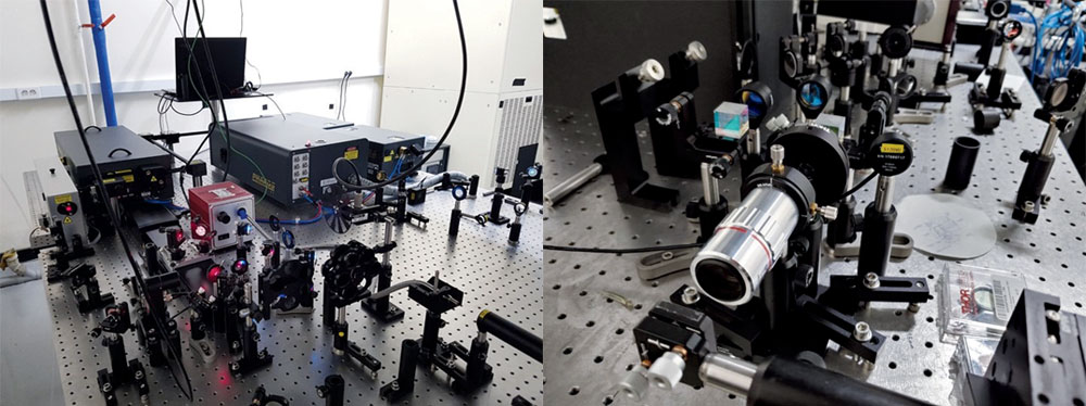
Figure 8. The femto-second laser systems and optics setups for time-resolved studies and non-linear optical experiments.
Ultrafast time-resolved optics investigates quasiparticle dynamics in a solid-state material using femto-second laser pulses, for understanding of how quasiparticles interact with each other to manifest collective phenomena and drive phase transitions. Our center is also actively working on ultrafast optical studies of various materials using our home-built optics setups, which consist of the femto-second laser system and optical parametric amplifiers installed in April 2019.
Our laser system can generate femto-second laser pulses over a wide range of wavelengths (from ultraviolet ~ 330 nm to far-infrared ~ 20,000 nm), which allows us to induce a diverse set of non-equilibrium states in a material and investigate their corresponding changes in the particle dynamics.
For example, wavelength- and polarization-dependent transient reflectivity studies can resolve the effects of heterointerfacing on the lifetime and stability of exciton particles. Additionally, using the same femto-second laser systems, we constructed non-linear optics setups for the symmetry analysis of materials.
The setup is based on a second-harmonic generation phenomenon, which has remarkable sensitivity to small changes in anisotropy and symmetry of a crystal, and has been actively used for discoveries of multipolar orders in various material phases. After completing a series of tests in normal-incident configurations, we are currently optimizing an experimental setup to effectively sample three-dimensional information of a crystal symmetry.
고휘도 엑스선 회절 장비는 물질 내 원자핵의 배열로부터 오는 격자 구조와 동역학을 밝히는 것에 초점을 두며, 전자의 동역학 연구에 초점이 맞추어져 있는 포항 가속기 내에 설치된 공명 비탄성 엑스선 산란 분광기와 상보적인 역할을 수행한다. 이러한 연구 목적의 달성을 위하여 강한 밝기의 엑스선 발생 장치와 높은 해상도의 엑스선 영역 검출기를 결합한 고휘도 엑스선 회절 장비를 현재 구축 중이다.
기존의 엑스선 발생장치는 출력을 높힐 경우 고체 상태의 타겟이 용융한다는 근본적인 한계를 가지고 있는데, 이 문제를 해결하기 위해 액체 상태의 타겟에 전자빔을 입사하는 새로운 방식의 엑스선 선원이 개발되었으며 이를 국내 최초로 도입하였다. 테스트를 통해 설계치인 80μm의 작은 엑스선 빔 사이즈와 8.7E11 ph/s/mm2의 높은 휘도를 확인하였다. 해당 장비의 안전한 운용을 위하여 납으로 차폐된 별도의 엑스선 실을 조성하였으며 자체적인 안전 연동장치 회로의 구축에도 성공하였다. 현재 자동화된 모터 테이블과 컨트롤 프로그램을 통해 장비의 자유로운 운용이 가능하다.
2020년 상반기에 고해상도 엑스선 영역 검출기가 구축이 되고 나면 본격적인 엑스선 회절 실험이 가능해질 것으로 기대가 된다. 단결정과 가루 형태의 시료에서 모두 결정 구조 분석이 가능해질 것이며, 영역 검출기의 장점을 활용하여 열확산신호를 통한 격자 동역학 연구도 진행될 예정이다. 열확산신호 데이터 해석을 위해 미국 Advanced Photon Source와 연구협력을 맺을 예정이며, 동시에 영역 검출기에서 생성되는 수 테라바이트에 해당하는 방대한 양의 데이터를 처리하기 위해 머신 러닝을 적용한 자체 분석 프로그램도 개발할 계획이다.
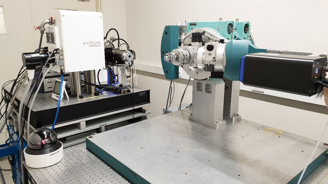
Figure 9. The high-brilliance x-ray diffractometer consisting of a metal-jet source and an image detector
The high-brilliance x-ray diffractometer is focus on the study of the lattice structure and dynamics of solids, complementary to the resonance inelastic x-ray scattering spectrometer (RIXS) installed in the Pohang Light Source focused on the dynamics of electrons in solids.
In order to achieve these research goals, a high-brilliance x-ray diffractometer that combines a high-brightness x-ray source and a high-resolution x-ray area detector is currently under development. Conventional x-ray sources have a fundamental limitation in increasing the power due to melting of the solid target.
To circumvent this problem, a new type of x-ray source that guide high-flux of electron beams electron beams to a liquid-state target was introduced for the first time in Korea. We confirmed that a small x-ray beam size of 80 μm and high luminance of 8.7E11 ph/s/mm2 can be achieved.
For the safe operation of the equipment, we constructed a separate x-ray room shielded with lead and implemented a safety interlock circuit system. Currently, the machine can be operated through an automated motor table and control program. It is expected that the x-ray diffractor will become operational once the high-resolution x-ray area detector is set up in the first half of 2020.
Crystal structure analysis will be possible for both single crystals and polycrystalline solids, and lattice dynamics studies through thermal diffusion signals will be implemented by taking advantage of the high-resolution area detector. To analyze the thermal diffusion scattering data, we will collaborate with Advanced Photon Source. Further, we also plan to develop a program that applies a machine-learning algorithm to process many terabytes of data generated by the area detector.
본 연구단에서 구축중인 극저온 고자기장 주사터널 현미경(그림 10)은 2019년도에 최종 조립을 마치고, 각종 테스트를 시행 중에 있다. 우선 냉동기 내부에 장착된 부품들의 접촉으로 발생한 열적 접촉을 제거하고, 또한 극저온 기저온도에 영향을 주는 복사열 차단 쉴드의 온도를 더 낮추어 냉동기의 성능을 향상 시켰다. 주사터널 현미경의 각종 기능을 담당할 전선 및 부품들을 장착하고도, 냉동기의 콜드 팁에 6 밀리켈빈 이하의 온도가 실현됨을 확인하였다. 또한 냉동기 내부의 각종 부품들도 주사터널 현미경의 사용에 편리하도록 수정 작업을 수행하였다.
냉동기 하부의 초고진공 챔버의 성능을 높이는 작업을 수행하였다. 우선 진공도를 높이기 위해, 4개의 액체질소 트랩을 새로 제작하여 추가하였고, 소형 이온펌프로 장착하여, 주사터널 현미경의 샘플 온도가 상온인 경우에도 1x10⁻¹⁰ Torr 수준의 초고진공도를 유지할 수 있게 하였다. 주사터널 현미경의 헤드를 냉동기 안의 초전도 자석 중심까지 이동 시킬 뿐만 아니라, 10 켈빈까지 샘플 온도를 낮추어 주사터널 현미경의 작동을 시킬 수 있는 매니퓰레이터의 최종 조립도 완성하였다. 냉각기능 및 전기 신호를 담당하는 60개의 전선을 탑재하였을 뿐만 아니라, 초고진공 챔버 내부을 볼 수 있는 내시경 3 세트도 탑재하여 기능을 극대화하려 노력하였다. 그림 11 참조.
2019년에는 이러한 각종 테스트를 수행하는 동안 발생한 여러 문제점을 해결하면서 기능을 향상 시키는 노력을 수행해왔으며, 2020년도 내에는 각종 저온 주사터널 현미경의 성능을 보여줄 수 있을 것으로 기대한다.
-
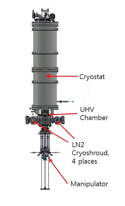
Figure 10. Newly Mounted Cryo-shrouds.
-
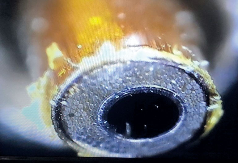
Figure 11. Endoscope End.
Our center’s Ultralow Temperature High Magnetic Field Scanning Tunneling Microscope (See Figure 10.) has been assembled, and tested in 2019. The performance of the cryostat has been improved by removing thermal contact between the critical parts inside of the cryostat and improving the temperature of the thermal shields, which is very crucial for base temperature of our STM and cryostat.
With these efforts, a base temperature of < 6 mK has been realized with all STM related parts and wires fully mounted in the cryostat. Some other important parts in the cryostat such as doors and shields has been modified or re-made to facilitate the STM operation in ultralow vacuum and low temperature.
We tried to improve the performance of the ultrahigh vacuum chamber which is beneath the cryostat. 4 sets of liquid nitrogen cryo-shrouds as well as 2 small ion pumps have been newly mounted to the chamber. With the help of these pumps, a vacuum base pressure of 1x10⁻¹⁰ Torr has been realized.
We assembled our STM head manipulator, which is a very crucial component of the whole system. The manipulator can cool down the sample as low as ~ 10 K, and enables operators to move the STM scanner into the center of the superconducting magnets in the cryostat. It has a cooling capability, and has 60 electrical wires for STM signals., as well as 3 sets of ultrahigh vacuum compatible endoscopes, with which operators can see the inside of the ultrahigh vacuum chamber and the cryostat. See Figure 11.
In 2019, we have tested the system, and tried to solve the problems which we found during various operation modes, and we expect we can show the power of this ultralow temperature and high magnetic field scanning tunneling microscope in 2020.
 Center for Artificial Low